
Mohs Micrographic surgery is the most effective treatment for skin cancer removal and where essentially 100% of the margin could be evaluated. It was named after Dr. Fredrick E. Mohs, MD who developed a technique called chemosurgery in the 1930s. A trainee of Dr. Mohs, named Dr. Perry Robbins, helped to advance the Mohs technique to what it is today.
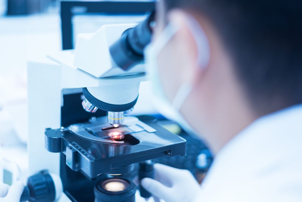
Mohs micrographic surgery drastically improved skin cancer treatment by its tissue sparing nature and higher cure rates. The treatment of skin cancer can generally be completed in one day under local anesthetic. The Mohs micrographic technique consists of removing the visible margin of skin cancer or left-over biopsy scar with a small margin of normal tissue. The removed tissue is then cut into sections, and colored coded to make a map. The tissue is then processed in the lab by a technician who places thin horizontal slices on a microscope slide. The Mohs surgeon will then be able to evaluate the margins of the tissue. If the cancer cells are seen on the slide, the Mohs surgeon will use the map to locate the positive margin and take another layer from the wound.
This process is repeated until all cancerous cells are cleared, then the wound is closed. During these steps the patient will wait in the office with a temporary dressing in place.
Mohs micrographic surgery is the gold standard for treating skin cancers such as basal cell carcinomas or squamous cells carcinomas in cosmetically sensitive areas such as the eye lids, lips, ears, fingers, toes, or genital skin. This technique can also be utilized for melanoma and other types of skin cancer as well. Drs. Sewell and Miner follow these applications of Mohs Micrographic Surgery as well as a modified version for superficial melanomas (the in situ type) as well.
Schedule a Skin Cancer Screening
If you have concerns with the health of your skin or need further information on Skin Cancer types, treatment, or prevention, please feel welcome to call High Valley Dermatology.